
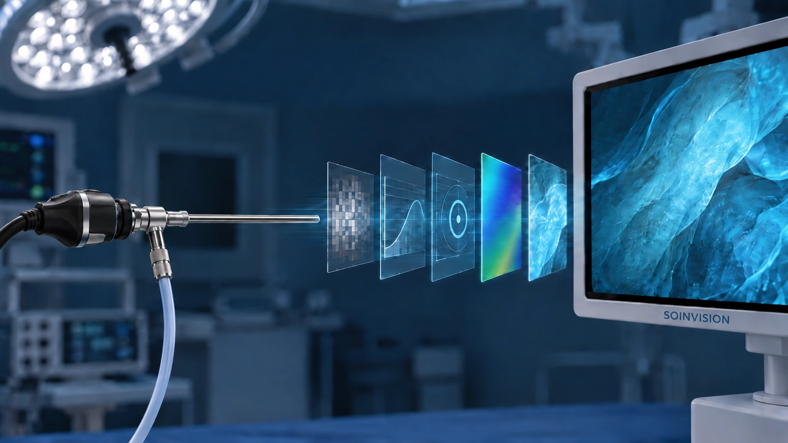
By the time video reaches a surgical monitor, it may already have undergone several upstream processing operations.
In an endoscopy workflow, video can pass through the camera system or camera control unit, a video output stage, additional downstream processing, and finally the display itself. Each stage can influence what appears on screen.
The useful question is not simply whether a system offers more enhancement. It is whether the existing video chain already performs the required processing, what visual problem remains, and where an additional processing stage would add value.
What Is Endoscopy Image Processing?
Endoscopy image processing refers to operations applied to a video signal or image data before or during presentation. Depending on the system, these operations may take place in the camera system, a dedicated processor, another device in the signal chain, or the surgical display itself.
Processing may adjust noise, tone, color, detail, resolution, or other characteristics of the video. Some operations prepare or condition the signal, while others are intended specifically to change how image information is presented.
Image enhancement is therefore better understood as one part of image processing rather than a synonym for the entire processing pipeline.
The distinction also matters when evaluating equipment. A display setting, a signal-management function, and an image-enhancement algorithm may all affect what appears on screen, but they do not necessarily perform the same role.
What Happens to Video Before It Reaches the Surgical Display?
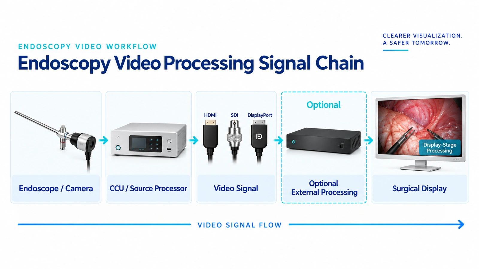
A simplified endoscopy video chain can be represented as:
Endoscope / camera → camera control unit or video processor → video output and transport → optional downstream processing → surgical display
The exact architecture varies by system. Some functions may be combined inside one device, while other workflows use several separate components.
Processing may already occur near the source. A camera system or video processor may apply operations such as white balance, gain adjustment, noise processing, tonal control, color processing, sharpening, or scaling before the signal leaves the device.
That means an additional processor or enhanced display does not begin with a completely untouched image. It receives whatever the upstream system has already produced.
This is an important starting point when evaluating whether another processing stage is useful.
What Types of Processing Are Common in Endoscopy Video?
Different systems use different processing pipelines, but several broad categories are common.
Noise Processing
Noise reduction can help suppress unwanted variation in the video image, particularly when signal conditions are difficult.
The trade-off is that stronger denoising can also reduce fine image texture or detail. The appropriate setting therefore depends on the source rather than on the assumption that more noise reduction is always preferable.
Tone and Contrast Processing
Tone and contrast processing changes how darker, mid-tone, and brighter regions are presented.
These adjustments can make some image information easier to distinguish, but aggressive settings can also compress or lose information in parts of the tonal range. The useful result depends on the source and viewing requirement.
Color Processing
Color processing may include white balance, color correction, saturation, color-space conversion, and related operations.
These functions should be separated conceptually from more specialized enhancement. Establishing the basic color presentation of the video and applying additional image enhancement are not necessarily the same processing step.
Detail and Edge Processing
Sharpening or edge enhancement can increase the apparent distinction around boundaries.
However, stronger enhancement is not automatically better. Research measuring edge enhancement in endoscopic systems found that increasing enhancement increased both measured sharpness and image noise.
That makes edge enhancement a useful example of why image-processing settings need to be evaluated as trade-offs rather than simply maximized.
Scaling and Resolution Adaptation
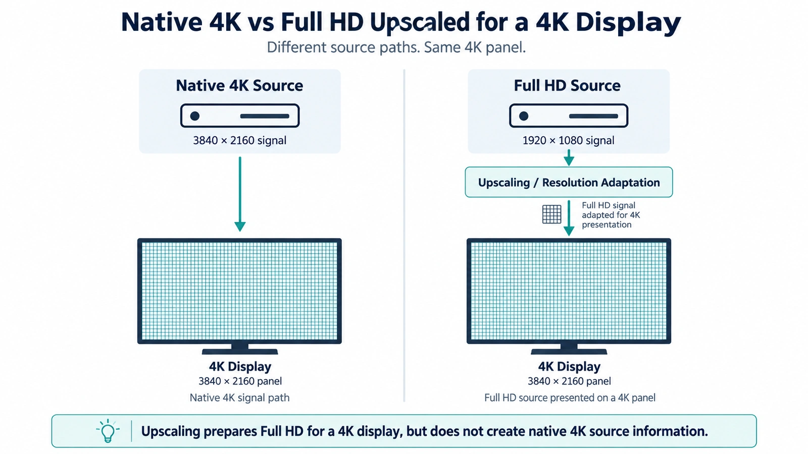
Processing may also adapt one video resolution for presentation on a display with another native resolution.
For example, a compatible Full HD source can be scaled for presentation on a 3840 × 2160 display. The resulting output can fill the 4K panel, but the operation does not turn the original Full HD acquisition into native 4K source information.
For a more detailed comparison, see native 4K and upscaled Full HD.
Compensation for Image Degradation
Some processing is intended to reduce the visible effect of image degradation such as haze or fog-like interference.
Digital defogging operates on the video image itself. Published research has evaluated processing methods for restoring visibility in foggy or smoky endoscopic video.
This should not be confused with physical surgical-smoke evacuation and filtration, which removes smoke or gas from the surgical environment rather than processing its appearance in the video signal. SAGES treats smoke and gas evacuation as a physical evacuation and filtration issue.
Why More Image Processing Is Not Always Better
A longer feature list does not automatically mean a better video result.
Every processing operation changes some part of the signal. Those changes can be useful, but they can also interact.
Stronger sharpening may make edges more pronounced while also making noise more visible. Denoising may suppress noise but remove fine texture. Contrast processing may improve separation in one tonal range while compressing another.
The same issue can arise when similar processing is applied at several stages.
If an upstream video processor already applies substantial sharpening, contrast adjustment, or noise processing, repeating aggressive processing downstream may produce a result that looks overprocessed rather than more informative.
This is one reason to evaluate the complete video chain rather than comparing isolated feature names.
Where possible, processing should also be evaluated against a neutral or bypass condition. Without a baseline, it is difficult to determine what a processing stage is actually changing.
Why Live Video Requires More Than a Good Still Image
A still frame can show whether an enhancement changes contrast, sharpness, or visibility at one moment. It does not show how the processing behaves over time.
Live video introduces another requirement: temporal consistency.
Processing that operates independently on each frame can produce changes that are more noticeable during motion than in a static image. Research into endoscopic video enhancement has specifically identified flicker as a problem that can occur when frame-by-frame enhancement does not adequately account for relationships between adjacent frames.
For this reason, evaluation should include movement, changing illumination, scene transitions, and other conditions found in real video—not just selected before-and-after screenshots.
Latency also belongs to the live-video evaluation.
Every device or processing stage can contribute to the overall signal path. Research into laparoscopic video delay has shown that added latency can affect performance and user experience under the conditions tested.
That does not establish one universal millisecond threshold for every endoscopy system. The relevant question is how the complete configured chain behaves under its intended workflow.
Where Should Additional Processing Happen?
If additional processing is needed, the next question is where it belongs in the video chain.
There are three broad possibilities.
Source-side processing takes place inside or close to the camera system or video processor. It is part of the video generated before downstream equipment receives the signal.
External processing adds a separate device between the source and downstream equipment. This makes the processing stage independent from the monitor and can be useful when an existing signal path needs an additional processing layer.
Display-stage processing takes place inside the surgical display. In this architecture, the display receives the incoming video and applies additional processing before the image reaches the panel.
None of these locations is automatically superior.
The right location depends on what the upstream system already does, what problem remains, how the equipment is being integrated, and whether the processed output needs to serve only the display or other downstream devices as well.
Do You Actually Need Another Processing Stage?
Sometimes the answer is no.
If the camera system already provides an appropriate image and the connected display presents it correctly, adding another processing device may provide little practical value.
Before adding another stage, it is useful to ask:
- What is the actual video source?
- What processing is already active upstream?
- What visual problem remains?
- Does the problem appear in the source itself, during transport, or only at the display?
- Can the new processing be compared against a bypass or neutral state?
- Will another stage duplicate processing that already exists?
- Does the project require processing for one display or for a broader downstream signal path?
This shifts the decision from “Which system has more enhancement features?” to “What does this particular video chain still need?”
Integrated Display Processing vs External Processing
Integrated display-stage processing and external processing can address similar visualization requirements while using different system architectures.
| Consideration | Integrated Display-Stage Processing | External Processing |
|---|---|---|
| Processing location | Inside the surgical display | Between the source and downstream display equipment |
| Hardware architecture | Processing and display combined | Processing is a separate device |
| Dependence on monitor | Tied to the display platform | Independent from the display |
| Existing-system upgrades | Add or change to an enhanced display | Insert processor into a compatible signal path |
| Additional hardware | Fewer separate devices | Adds another device and connection stage |
| Downstream signal use | Primarily display-centered | Can support downstream equipment where appropriate outputs and routing are available |
| Typical project context | New or display-centered systems | Retrofit, mixed-system or independent-processing projects |
This is an architecture choice rather than a quality ranking.
An integrated approach may simplify a system in which enhancement is primarily needed at the main display. An external approach may provide more flexibility when processing needs to remain independent from a specific monitor or when an existing system is being upgraded.
How to Evaluate Image Processing in a Real Endoscopy Workflow
Feature lists are useful for establishing what a device can do, but they do not show whether a particular processing setup is appropriate for a real signal chain.
Evaluation should begin with the actual source that will be used in the system.
Check the source resolution, signal format, video output, and the processing already being applied. If possible, compare the additional processing with a neutral or bypass state rather than relying only on demonstration images.
Then evaluate the areas that matter for live use:
Image behavior — Does the processing improve the specific issue being evaluated without introducing distracting noise, halos, excessive contrast, or an unnatural appearance?
Adjustment range — Can processing strength be reduced or disabled when the source does not need the same level of enhancement?
Temporal behavior — Does the result remain stable as the image moves or lighting conditions change?
Latency — Does the complete video path remain suitable when all processing and transport stages are active?
Integration — Do the input, output, resolution, and routing requirements match the surrounding equipment?
Downstream use — Is the processed signal needed only on one display, or must it also feed another monitor, recorder, router, or other equipment?
When image-processing requirements are part of a wider display project, these checks should also be considered alongside the other factors involved in choosing a 4K surgical monitor.
Testing with the real source and intended workflow is more informative than choosing a system from an isolated enhancement demo.
How SOINVISION Implements These Two Processing Architectures
SOINVISION uses both of the processing architectures described above, but they should not be treated as the same product concept.
In the NEXARA X series, S-VIXTA image enhancement is integrated into the surgical display. This is an example of display-stage processing: enhancement is part of the display platform rather than a separate processor inserted into the signal chain.
S-VIXTA adds functions including Global Optimize, Defog, Depth Enhancement, Vessel & Tissue Visibility Enhancement, and adjustable enhancement intensity to the shared NEXARA display platform.
MILESTONE follows the external-processing approach. It is a separate medical video image-processing platform that can be placed in a compatible signal path before downstream display equipment.
The distinction is primarily architectural:
NEXARA X + S-VIXTA → processing integrated at the display stage.
MILESTONE → processing provided by an independent external platform.
Which approach is more appropriate depends on the existing video chain and project requirements rather than on the assumption that one architecture is universally better.
Start With the Video Chain, Not the Feature List
Endoscopy image processing is most useful when it addresses a specific requirement in the video chain.
The source may already perform substantial processing. A downstream processor may be useful in an existing system that needs an independent enhancement stage. A surgical display with integrated processing may make more sense when the enhancement is intended primarily for the final viewing stage.
The same principle applies to individual processing functions. Sharpening, denoising, contrast adjustment, scaling, defogging, and other operations can all be useful, but their value depends on the source, the surrounding processing stages, and how they behave in live video.
Before choosing additional processing, identify what the system already does, what problem remains, and where that problem can be addressed most appropriately.
That provides a more reliable basis for evaluating an endoscopy visualization workflow than comparing feature counts alone.